NTA is a technique used to measure the size and concentration of exosomes based on the analysis of Brownian motion. The scattered laser light of the particles is recorded with a camera. Molecular diffusion is determined by tracking changes in a single particle set and then converted to a hydrodynamic diameter (using the Stokes-Einstein equation).
Importance of exosomes in cancer proliferation and diagnoses
Exosomes are spherically shaped extracellular vesicles (EVs) with diameter within the range of 30-150 nm bounded by a lipid bilayer, secreted from many human cells. They reveal the big importance in long-distance intracellular communications and propagation of diseases, especially cancer. Many cancer cells produce an increased amount of exosomes in comparison to healthy ones. It can be supposed that one can recognize what type of cell produced the given exosome and what was the health-state of its cell of origin, basing on the contents (miRNAs, mRNAs and proteins).Exosomes can be found in body fluids like blood, saliva or urine and can be used as biomarkers of specific cancer. In the case of prostate cancer it might be of great importance, because it is not easy to diagnose it, for various reasons, and noninvasive screening methods are in demand.
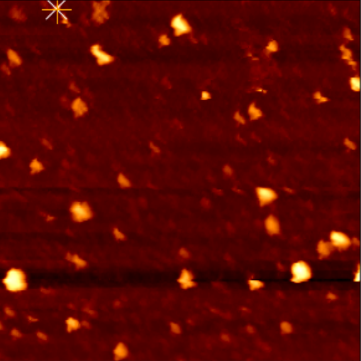
AFM image of exosomes deposited on mica surface pre-modified with APTES
Scientific goals of the project
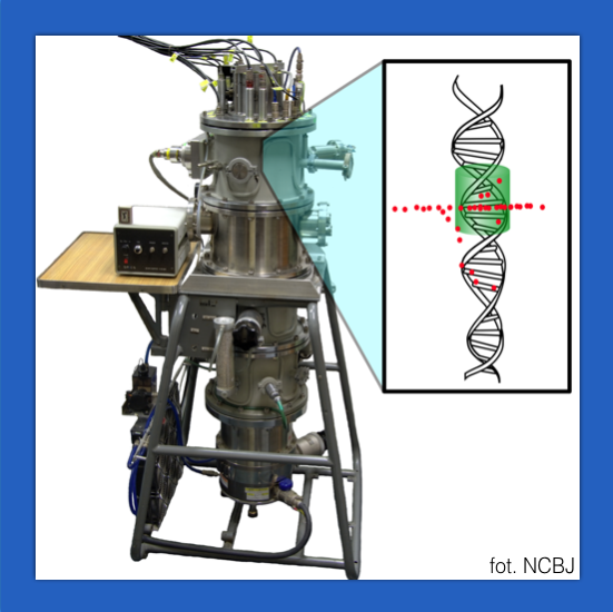
The first goal of this project is to investigate in detail the biophysical properties of exosomes derived from prostate cancerous and healthy cells. The hypothesis to be verified is that microscopic methods such as NTA (Nanoparticle Tracking Analysis) and AFM (Atomic Force Microscopy) provide a possibility to identify and distinguish sub-populations in prostate derived exosomes. The latter method enables the imaging of 3D shapes of single objects at the nanoscale. It can provide information not only about their size and morphology but also about the adhesion, elasticity, and deformability of extracellular vesicles.It has been demonstrated that cancer cells often defend themselves against radiotherapy. Exosomes secreted from cancer cells treated by ionising radiation revealed the changed amount and contents that can affect exosome-based intercellular communication and promote radioresistance. The second hypothesis to be verified is that properties of exosomes isolated from prostate cancer line cells irradiated with ionizing alpha or beta radiation might be distinguished from the ones derived from non-irradiated cells employing NTA and AFM based methods. Examination of properties of radiation derived exosomes could be a tool of prognosis in radiotherapy treatment. The third goal of the project is to review the exosomes’ effects on prostate cancer progression, especially to the tumorigenesis and metastasis.
Results
1. Methodology of cells culture (commercial lines PC3 and DU145), alpha irradiation and exosomes isolation2. NTA based confirmation of exosomes presence, measurements of their size and concentration
3. Methodology of mica substrate preparation and AFM imaging of nanovesicles
4. AFM based analysis of size and biophysical properties of prostate cancer exosomes
- Beata Brzozowska
- Józef Ginter
- Wioletta Olejarz
- Tomasz Lorenc
- Katarzyna Życieńska
- Adrianna Tartas
- Mateusz Filipek
- Beata Pszczółkowska
The studies are performed within cooperation between:
- Faculty of Physics and Faculty of Chemistry (University of Warsaw)
- Faculty of Pharmacy and Faculty of Medicine (Medical University of Warsaw)
Partial financing: funds for cooperation between WUM-UW (Polish title: Badanie wpływu promieniowania jonizującego na profil egzosomów pochodzących z komórek raka prostaty o różnej promieniowrażliwości; dr G. Kubiak-Tomaszewska, mgr A. Tartas)
The project’s goal is to investigate the biophysical properties of exosomes isolated from irradiated and non-irradiated prostate cancer cells employing NTA and AFM based methods. We try also to review the exosomes’ effects on prostate cancer progression, especially to the tumorigenesis and metastasis.