
The aim of radiation therapy is to deliver the prescribed dose of ionizing radiation to the target volume without increasing normal tissue complications. The dose distributions calculated in the treatment planning system are depended on many parameters. One of them is the shape of PTV (Planning Target Volume) which is drawn by the medical doctors and contour of OARs (Organs At Risk) delineated by medical physicists and electroradiologists. The goal of this project is to investigate intra- and inter-observer variability in contouring target volume and organs at risk and its impact on conformal treatment planning. The project assumes statistical and geometric analysis the contours which are manually inserted by physicians and medical physicists. Measured and calculated dose distributions will be compared (based on DVH histograms and descriptive statistics) for different cancer localization using the PlanUNC treatment planning system. The project is divided in two parts, which will be investigated independently.
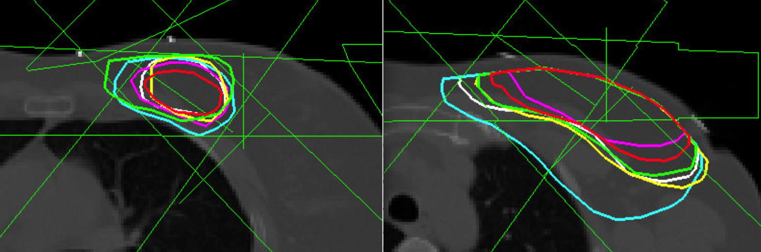
In the FIRST PART six oncologists contoured the PTV and boost regions for a breast cancer patient. The main purpose was to compare the contours using their geometric fields (the volume of each contour, centroid and the percentile of sharing field). The influence of contouring for the dose in PTV region is examined. The PTV parameters (V90% and V95%) were estimated and plotted as a function of the total target volume.
In the FIRST PART six oncologists contoured the PTV and boost regions for a breast cancer patient. The main purpose was to compare the contours using their geometric fields (the volume of each contour, centroid and the percentile of sharing field). The influence of contouring for the dose in PTV region is examined. The PTV parameters (V90% and V95%) were estimated and plotted as a function of the total target volume.
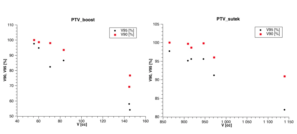
The target volume differs significantly dependently on the contouring physician and more studies are needed.
In the SECOND PART we will analyze the contours of the organs at risk which will be drawn by medical physicists. There will be intra- and inter-observer analysis. Firstly the medical physicists will contour the same area day by day for the whole week. Secondly they will contour few different areas for 10 patient plans. In this case the analyze will be based on DVH histograms. The maximum dose, minimum dose, median dose and mean dose in each organ at risk will be found. Also the standard deviation of the dose will be calculated. The geometric analysis will be the same as in the first part of the project.
The project is a result of cooperation between Faculty Of Physics, University of Warsaw (represented by Student Union), Maria Skłodowska Curie Memorial Cancer Centre and Institute of Oncology and Greater Poland Cancer Centre.
OUR TEAM
- Anna Zawadzka, project co-leader (Maria Sklodowska-Curie Memorial Cancer Center)
- Beata Brzozowska, project co-leader (University of Warsaw)
- Bartosz Bąk (Wielkopolskie Centrum Onkologii)
- Zofia Biały (University of Warsaw)
- Edyta Dąbrowska-Szewczyk (University of Warsaw)
- Józef Ginter (University of Warsaw)
- Adrianna Tartas (University of Warsaw)
Only in Polish: Raport Zespołowego Projektu Studenckiego