
The mechanisms of consciousness are among the greatest mysteries of neuroscience. Research within the area of disorders of consciousness (DoC) aims to broaden our knowledge of the theoretical basis of these processes and to provide diagnosis-supporting solutions for healthcare. Studies conducted so far have shown promising results in applying neuroimaging techniques such as nuclear magnetic resonance (NMR) and electroencephalography (EEG). However, recent applications of both methods in assessing DoC severity leave room for significant improvements in signal processing methods, which leads to suboptimal sensitivity and specificity. Furthermore, there is a lack of longitudinal studies combining these two methods while evaluating the patient’s state during the DOC evolution from the very beginning, i.e., from the state of coma. The project’s main interest is to fill these gaps within the contemporary research on DoC.
The longitudinal protocol of measurements for each patient will consist of the following steps: an examination immediately after the accident, after approximately two weeks since the accident (both measurements conducted during the state of coma), after the awakening from coma (in a vegetative state), minimally conscious state and after the complete recovery of consciousness. Biochemical changes in the brainstem will be monitored using magnetic resonance spectroscopy. The formation of new functional connections will be assessed based on the results of resting-state activity in a magnetic resonance (MR) scanner and changes in brain functioning—via observation of resting-state electrical brain activity. A reference for the neuroimaging methods described above will be provided by a clinical scale specific to monitoring the recovery of consciousness (Coma Recovery Scale-Revised, CRS-R), supplemented by EEG-based indicators developed as a part of the NCN project “Brian/neural computer interaction for diagnosis and communication in disorders of consciousness”. Analysis of data provided by both of the neuroimaging methods mentioned above (MRI and DOC) require significant improvements for an effective application in DoC research; hence the development and validation of novel algorithms are planned within the Project.
EEG-based studies of coma are traditionally based on visual detection of characteristic structures present in resting-state EEG recordings, such as triphasic waves, delta activity, coma spindles, and K-complexes. A similar approach, applied to the analysis of circadian activity (presence of which, according to several studies, may be an important indicator of the patient’s progress) is based onhypnograms. Such evaluation is classically performed by visual inspection of raw EEG recording, which negatively influences repeatability and costs of the procedure and sensitivity of proposed indicators. The Applicant has several years of experience in the automated detection of structures present in sleep EEG (such as e.g.spindles, slow waves, K-complexes), based on their definitions used in visual analysis and in the construction of hypnograms based explicitly on the occurrences of automatically detected structures. Approaches published by the Applicant, developed for standard hypnograms, were also applied to the parametrization of sleep of patients with DoC, first in cooperation with prof. Laurey’s clinic in Liege, and then as a part of the NCN project “Identification of electroencephalographic patterns in recordings of spontaneous brain activity in children with disorders of consciousness”. Results of these studies, published in prestigious journals, point the way to the automation of the assessment of the occurrence of circadian rhythmicity, which could be based on selective detection of relevant structures with parameters from time-frequency space, obtained by decomposing the signal with the matching pursuit (MP) method, introduced to the analysis of biomedical signals by the Applicant.
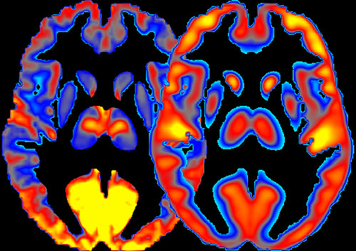
An important extension of the proposed methodology is the application of structural (MRI) and functional (rsMRI)Magnetic Resonance Imaging. sMRI offers an improved description of a patient’s structural brains damages, while rsMRI allows identifying a network of “cooperating” areas in the brain, known as the default mode network (DMN). MR signal in DMN areas results from the convolution of neuronal activity with hemodynamic response function (HRF). HRF response covers a cascade of metabolic processes such as oxygen extraction or regulation of brain perfusion. Therefore, an important part of the project will include the development of a new method of rsMRI signal analysis, taking into account the spatial variability of HRF. Finally, single-voxel MR Spectroscopy (svMRS) will be used to measure selected brain metabolites (esp. neurotransmitters) since their concentration may provide indicators related to the changes in the level of consciousness.