Dose is a macroscopic quantity defined as the energy absorbed per unit mass. It is one of the fundamental concepts in nuclear and medical physics and plays a central role in radiotherapy planning. Modern radiotherapy aims to deliver the prescribed dose to the target tumor volume while minimizing irradiation of surrounding healthy tissues. Although photon beams remain the most widely used modality, proton and light-ion therapies are increasingly adopted due to their favorable depth-dose distributions and enhanced radiobiological effectiveness.
While the effects of ionizing radiation are mostly dependent on the absorbed dose, direct comparisons between different radiation qualities are not straightforward. To address this limitation, a dose-related quantity known as the relative biological effectiveness (RBE) was introduced. RBE is defined as the ratio of the absorbed dose of a reference radiation to that of the radiation under investigation required to produce the same biological endpoint, such as a given level of cell survival.
To understand why different types of radiation have different biological effectiveness, let us use the example presented in figure 1. The difference lies in how radiation interacts with cell nuclei. If one imagines a specific absorbed dose administered by photons to a macroscopic volume the interactions will be nearly homogeneously distributed across the target volume. As a result, a large number of sensitive targets are affected, but each interaction typically induces relatively simple damage that can be efficiently repaired by cellular repair mechanisms. In contrast, heavy charged particles produce highly inhomogeneous and the majority of sensitive DNA sites will not be traversed, but some sites will be severely damaged.
At the cellular level, a more critical form of DNA damage is the double-strand break (DSB), as it is considerably more difficult to repair than a single-strand break (SSB). DSBs occur more frequently following irradiation with heavy ions, whereas SSBs are more typical for photon radiation.
In result, cell survival probability depends not only on the absorbed dose but also on the structure of the particle tracks. However, the DNA molecule has nanometric dimensions, far smaller than the typical ranges of secondary particles produced during irradiation. This means that dose and RBE, as the macroscopic quantities, is not the most suitable measure to describe DNA damage at the molecular level.
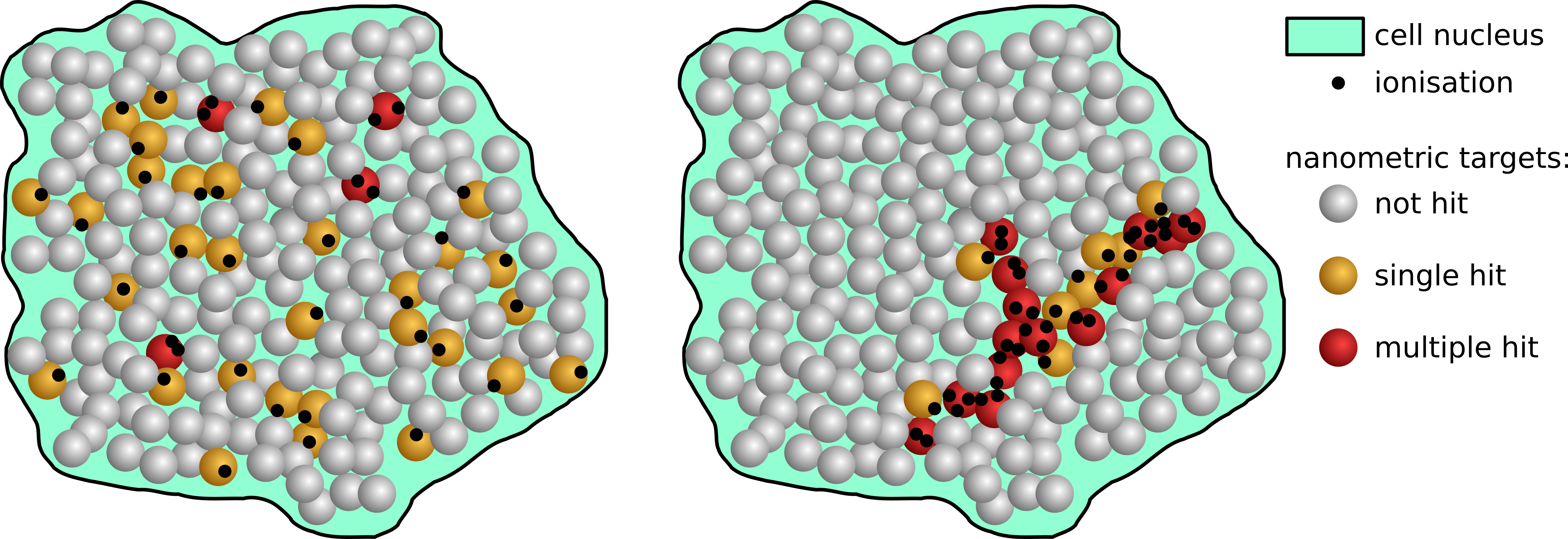 Figure 1: Ionisation patterns in cell nucleus.
This is where nanodosimetry comes into play – a field dedicated to characterizing how ionizing radiation interacts with matter at the nanoscale. It focuses on the stochastic nature of radiation interactions within a nanometric, most often cylindrical, volume equivalent to DNA segments and investigates the track structure of ionizing radiation. A key concept in this field is the ionization cluster size distribution (ICSD), which quantifies the number of ionizations produced within a nanometric target volume by a single particle track.
Both experimental and numerical techniques have been developed to study nanodosimetry. One of the most important numerical approaches is Monte Carlo (MC) simulation. They are based on defining a sensitive target volume and simulating tracks of individual particles. On this basis, ICSD probabilities are determined. These simulation codes offer high accuracy and are developed using specialized frameworks such as Geant4-DNA or TOPAS-nBio.
A major advancement in the field was the development of the nanodosimeter detector. It is based on a low pressure gas system that simulates nanometric biological targets. The device is capable of measuring ionisation clusters for single heavy charged particles. The number of such events allows for estimation of ICSD. At present, those detectors are the only experimental nanodosimetry technique, capable of modeling DNA damage with sufficient resolution. One of them is Jet Counter nanodosimeter developed at the National Centre for Nuclear Research
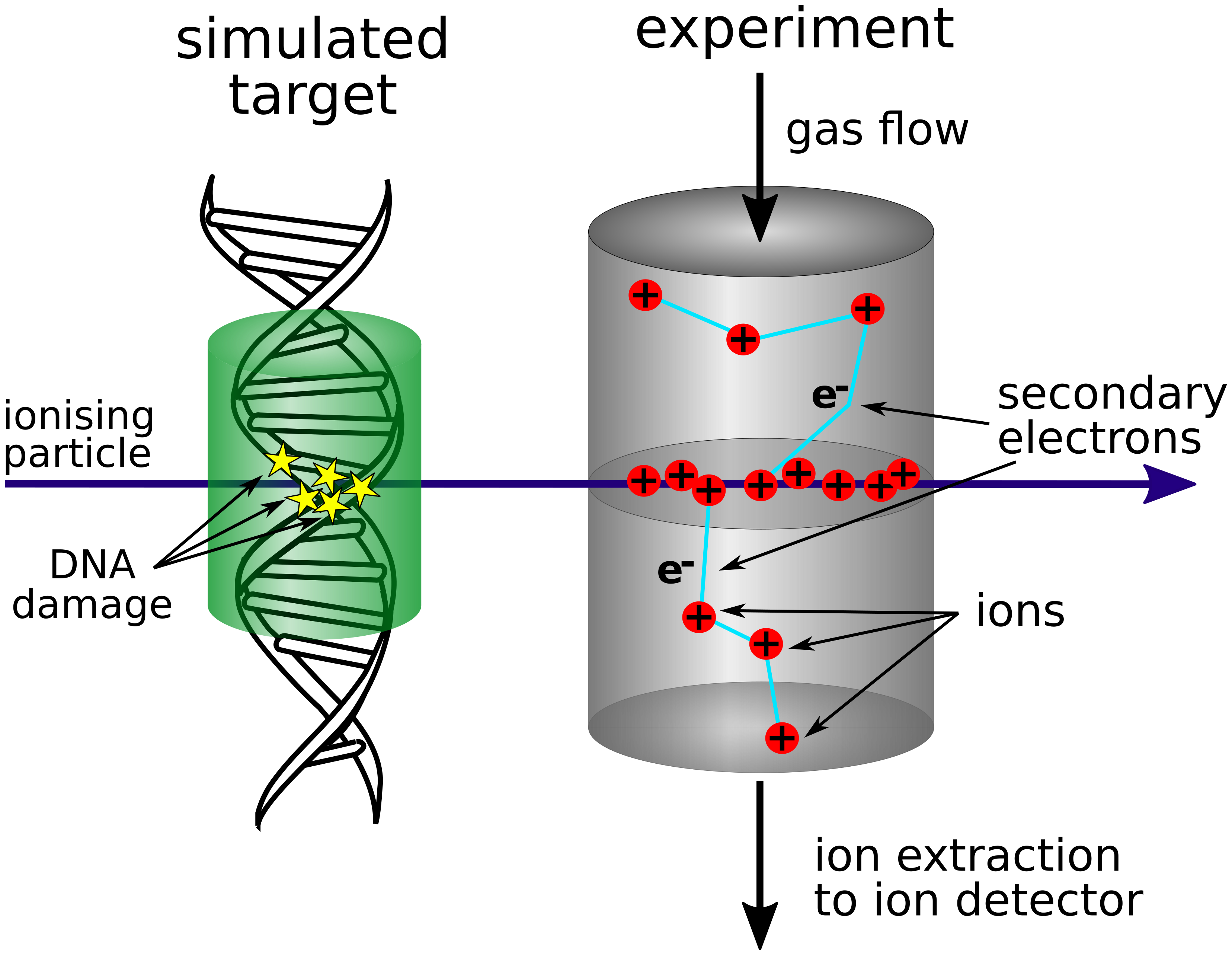 Figure 2: Schematic view of simulated nanometric target and its experimental realisation.
On the one hand, experimental results can be used as excellent benchmarks for simulations, on the other, simulations allow obtaining information that is not available by experimental methods.
One of the key challenges is establishing a quantitative link between radiobiological dose measurements and nanodosimetric parameters. Several approaches have been proposed to address this problem. One of them is the Fk parameter, which describes the probability of inducing k or more DNA damage events within a sensitive volume. An alternative approach proposed by our group is the R₂ parameter (equation 1, where P𝜈 is a probability distribution of cluster size 𝜈, Bk (𝜈, p) is the probability of k ionizations that have provided a sub-lethal lesion in a sequence of 𝜈 ionizations, and p is the probability of creating a sub-lethal lesion by a single ionization in this sequence). While conceptually similar to F2, R₂ additionally accounts for the probability of sublethal DNA damage. The R₂ parameter can be directly related to the formation of DNA double-strand breaks, providing a more comprehensive description of radiation-induced damage at the nanoscale. These parameters have previously been evaluated using an online radiobiology database and Monte Carlo simulations.

Eqn 1: R2 parameters proposed by the group.
The aim of the current project is to investigate radiobiological responses in controlled experimental setups and to compare the results with nanodosimetric parameters from simulations. Experimental studies will include measurements performed on plasmid DNA as well as on cancer cell lines.
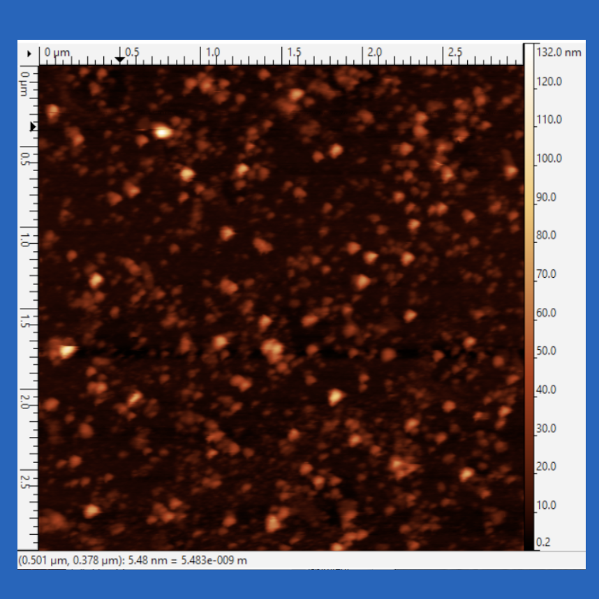
Plasmid DNA is a small, circular molecule of double-stranded DNA that exists independently of the chromosomal DNA in bacteria. Plasmids are commonly used in biology and radiobiology experiments because they offer a simplified model for studying DNA damage and repair mechanisms. Plasmid pUC19 will be irradiated in an experimental setup. Following irradiation, the plasmids will be analyzed from two perspectives: nanodosimetry analysis based on Monte Carlo simulation and biological assessment. The biological analysis will include an electrophoresis method and AFM analysis.
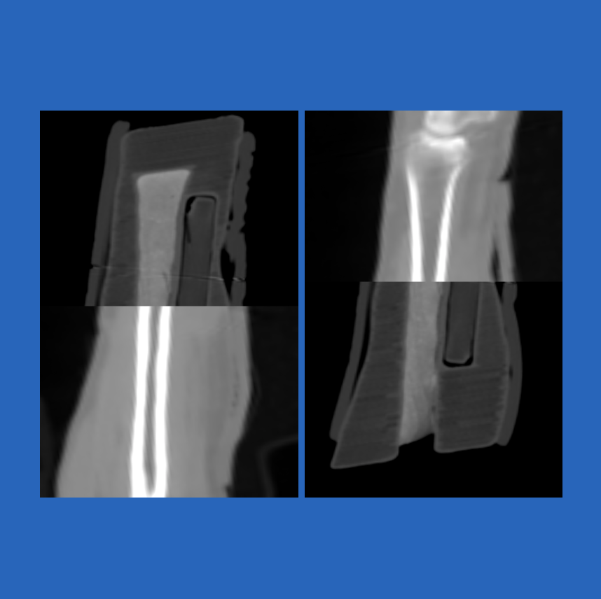
The next part of the experiment will be conducted using the human osteosarcoma cell line U2OS. This cell line is widely used in DNA damage and repair research because of its well-characterized and reproducible response to ionizing radiation. The cells will be irradiated under the same conditions as the plasmid DNA. Nanodosimetric analysis will follow the same approach as for the plasmid DNA. Biological analysis, which will be performed using the γH2AX assay.
OUR TEAM (alphabetically)
- Aleksandr Bantsar (IFJ PAN)
- Beata Brzozowska (University of Warsaw)
- Mateusz Filipek (University of Warsaw)
- Piotr Gasik (GSI)
- Monika Kopińska (University of Warsaw, NCBJ)
- Victor Merza (Universidade de Lisboa)
- Marcin Pietrzak (IFJ PAN)
- Antoni Ruciński (IFJ PAN)
- Stanisław Pszona (NCBJ)
- Zygmunt Szefliński (HIL)
- Agata Taranienko (University of Warsaw)